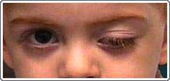
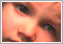
Child with left third nerve palsy after a virus. The left eyelid is droopy (ptotic), and the left eye is slightly down and out when the right eye looks straight ahead.
Muscle palsies are diagnosed by the inability of the eye to move as it should. Special tests are performed to see how the eye moves in different directions. Diagnosis of muscle palsy in children is especially difficult. Palsies might present as isolated events or be associated with other neurological problems. If accompanied by other neurological signs, the patient should have a neurology evaluation.
Third Nerve Palsy
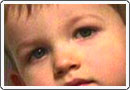
The third nerve innervates four of the six extraocular muscles as well as the muscle that elevates the upper eyelid. That is why a third nerve palsy is associated with weakness of several eye muscles; this makes it very difficult to treat. In childhood, a third nerve palsy is typically accompanied by other neurological findings, which helps in the diagnosis. But isolated palsies do occur and are generally congenital, traumatic, infectious, or secondary to migraines (yes, children can have migraines!). An acquired isolated oculomotor nerve palsy in a child may also result from tumor, preceding viral illness, bacterial meningitis or even immunization. Non-traumatic third nerve palsy cases must undergo a full workups with neuro-imaging.
The usual clinical sign of a third nerve palsy is the outward and downward location of the involved eye. This may or may not be accompanied by lid drooping. The pupil might also be larger when compared to the other side. The treatment of third nerve palsy is complicated since more than one muscle is usually involved. Usually a period of observation is prescribed to see if it resolves spontaneously before surgery is considered. The main goal of surgery is to move the eye into its neutral position (looking straight ahead) because no surgery can strengthen the already paretic muscles. Eyelid surgery is not recommended until after the eye muscle problem is taken care of.
Fourth Nerve Palsy
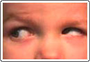
Commonly referred to as superior oblique palsy, this is a very common type of palsy seen in children and adults. Of all the causes of fourth nerve palsies in children, congenital and traumatic are by far the most common. It is usually unilateral, and patients present with facial asymmetry and a head tilt to one side to avoid double vision. In children, parents or primary physicians often notice the head tilt first and then seek consultation.
In congenital cases with a head tilt, surgery is usually planned as soon as the diagnosis has been established to prevent the development of facial asymmetry, abnormal neck muscles, and misalignment of spine. However, fourth nerve palsy can often go undetected until late adulthood when the patients start complaining of double vision, especially when tired. Acquired fourth nerve palsy develops secondary to head trauma, and is often associated with double vision and patient discomfort. Both eyes may be affected with a head trauma. A waiting period of 6-12 months is advised to observe for spontaneous resolution of the palsy before any surgery is recommended.
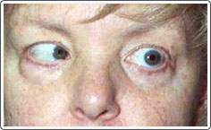
Young Child with Congenital Fourth Nerve Palsy
Patient adopts a compensatory head tilt to the right to keep the eyes aligned
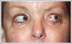
Left inferior oblique muscle over action caused by a weak left superior oblique muscle. Note the left eye is elevated.
With forced head tilted to the left there is a left hypertropia (left eye elevated) which is not comfortable for the patient.
Post-operative photo showing corrected head position and eyes are well-aligned in primary position after eye muscle surgery.
Sixth Nerve Palsy
Sixth nerve palsy is associated with a limited ability of the eye to turn out. The patient with a sixth nerve palsy often develops a head turn to get rid of the double vision and maintain binocular fusion. It is usually unilateral.
A congenital sixth nerve palsy is extremely rare; however, some newborns may have a sixth nerve palsy that resolves spontaneously over a few days to a few weeks. In early childhood, a common cause of isolated acquired sixth nerve palsy may occur 1 to 3 weeks after a viral illness or immunization, or may occur spontaneously without obvious cause. These patients should be followed closely to monitor their improvement and watch for the development of amblyopia. Improvement usually occurs within 6 to 10 weeks. Closed head trauma or intracranial lesions can also cause acquired sixth nerve palsy. Intracranial tumors should always be considered in cases of sixth nerve palsies. An MRI is indicated if the palsy does not improve rapidly, or if other neurological signs are present. Other causes of acquired sixth nerve palsy include meningitis, myasthenia gravis, and cavernous sinus disease, mostly seen in adults.
Patient with left sixth nerve palsy. Pre-operative photo shows inability of the left eye to turn out in left gaze.
Post-operative photo shows recovered ability for the left eye to turn out in left gaze.
Initial therapy of sixth nerve palsy is observation for 6 months while monitoring the patient for spontaneous recovery, approximately 80% for unilateral cases and 40% for bilateral cases. During the observation period, covering the eye with a type of patch or using press-on prisms can help to eliminate double vision. After the six-month observation period, the doctor will evaluate the patient for further treatment including possible surgery or prism glasses. Sometimes doctors use botulinum to inject into the affected muscle, paralyzing the muscle for 3 to 6 months. The hope is that preventing secondary contracture of the medial rectus muscle will increase the chances of recovery without strabismus surgery. The use of botulinum for treating sixth nerve palsy remains controversial and its effectiveness has not been proven.