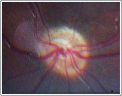
Through fundus photography, we are able to see the optic nerve in the back of the eye. In patients with optic nerve hypoplasia, the rim of the nerve is blurred and extended out onto the retina. The size of the center is smaller than normal.
Optic nerve hypoplasia is a non-progressive congenital problem affecting one (unilateral) or both (bilateral) optic nerves. The optic nerve is the pathway from the brain to the eye that allows interpretation of vision to take place in the brain. It is the most common developmental anomaly of the optic nerve and may be associated with multiple central nervous system and endocrine disorders. The endocrine abnormalities associated with optic nerve hypoplasia are treatable, and an endocrine work-up should be obtained on patients with this optic nerve disorder.
Optic nerve hypoplasia may be sporadic; however, other factors have been implicated including the use of drugs such as quinine, lysergic-acid-diethylamide (LSD), phencyclidine (PCP), alcohol (50% of fetal alcohol syndrome); intrauterine infection; cytomegalovirus (CMV); and possibly maternal diabetes. Optic nerve hypoplasia is bilateral in 60% of all cases; however, when bilateral, it often presents with nystagmus and neonatal blindness. Unilateral cases usually present with strabismus, as the eye with the optic nerve hypoplasia tends to drift causing either esotropia (inward turning) or exotropia (outward turning).
Visual potential does not necessary correlate with the size of the optic disc, and there is no specific treatment for optic nerve hypoplasia.